What is a hollow foot?
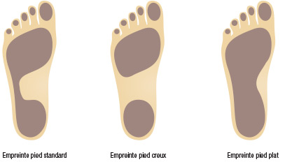
It is also referred to as a “cambered foot” or “arched” foot, and presents itself with an accentuated plantar arch (or internal arch). The foot appears as if the sole of the foot had been dug.
In reality it is mainly the footprint that determines the definition. To appreciate it, you must use a podograph or a podoscope. The podograph makes it possible to mark by the passage of a layer of ink on a sheet of paper, the imprint of the foot.
Clinically, a hollow foot has an exaggerated camber, sometimes with a bum on the sole of the foot, under the head of the big toe, or with a metatarsal head of the big toe lower than the other toes, visible especially when examining the front foot. It is often accompanied by a varus of the rear foot and / or a first metatarsal dip. With wear and / or characteristic deformation of the shoes. In fact, it is the reverse of a flat foot.
Sur un podoscope, son empreinte est réduite aux zones d’appui antérieur et postérieur. Habituellement ces 2 triangles d’appui sont réunies par une zone d’appui intermédiaire dite «isthme». Dans le cas du P.C. et selon la largeur de l’isthme, on distingue, 1er, 2e et 3e degré.


Hollow foot
Q & A
Do you need X – Rays?
If there is no pain or difficulty in heating, however if an X-Ray report is requested, it can show some signs. For the architecture of the foot is preserved in the rough cheeses or those of the first degree. But in the advanced forms, or in those of the adult, it may show a “break” of the line of the first ray which unites the following bones: talus, navicular bone, 1st cuneiform, and 1st metatarsal, with an angle at the apex Upper or back. The hollow foot is in the child and the young adult, not or little symptomatic. Over the years, a dilemma with the shoe, appears, especially with high heels in women. Pains occur at the level of the plant or at the dorsal level of the foot: at the mid-tarsus. They may be accompanied by signs of mid-tarsal osteoarthritis visible on the profile Xray: pinching of the articular interlining; Formation of osteophytis . The back foot is often supinated and to compensate for, the forefoot is pronated. The result is the onset of the following symptoms:
- Cramps and often dial heel,
- Metatarsalgia and very often instability of the ankle,
- Retraction of the extensors and sometimes grime of the toes,
- Plantar callus and episodically pain under the heel: talalgia.
Additional examinations supplement, if necessary, clinical and podoscopic examination: x-rays, EMG and / or MRI. A Neurological etiology symptoms may accompany a hollow foot, the presence of other signs may require the opinion of neurologist.
How do we treat Hollow feet?
If there are no other signs or if there has no grievances, there is no treatment. Simply supervised by the podiatrist or the pediatrician or the attending physician. If there is an embarrassment or a real pain and after eliminating a neurological cause, the treatment is essentially medical and is based on a triptych:
- Footwear: a good footwear, low heels often relieve and su sens.
- Soles: which allows the surface to be spread out and to reduce the pressure, (Pressure = Weight / surface, if the surface increases the pressure decreases).
- Rehabilitation: In some cases, stretching exercises in self-rehabilitation can reduce cramping especially if they are associated with good footing and / or a good footing.
In the majority of cases: The wearing of foot orthoses can solve the problem of cramps and pain. Self-rehabilitation (stretching exercises) completes the therapeutic range. Surgery is the treatment for severe cases.
Is it necessary to operate the Hollow Foot?
In general no, but there are forms linked to causes that require it. In severe or rebellious forms of medical treatment, surgery may be required. It is based on the nature of the distortion and its importance. In which cases ? And which kind of surgical procedures ? The indications can range from a simple plantar aponeurotomy (soft tissue procedure) to abone procedure including or not an articular procedure) like :
1. Osteotomy for raising the first metatarsal: This is an intervention that raises the head of the 1st metatarsal and reduces the pressure from under the head of the first radius. There is often a bone fixation, screw, staple, or temporary or permanent pin.
2. Release of the soft tissue to the medial side of the heel: In certain severe forms or neurological origin, with spasticity, a gesture on the soft parts becomes indispensable.
3. Osteotomy of the calcaneus (Dwyer’s operation): This is an intervention which makes it possible to straighten the hindfoot to increase the lateral support, especially if the hindfoot is in exaggerated valgus> 10 °, And which requires fastening with screws or staples.

4. Tarsectomy: This is a slightly heavier gesture because it involves a partial resection of the tarsus, in particular to remove this break from the midfoot and align the first ray,
5. Mid-tarsal arthrodesis consists of blocking a joint by correcting the axial deflection, it eliminates part of the mobility of the foot and reduces or even suppresses the pain and allows the foot to be aligned while retaining the mobility of the ankle and the toes.
6. Cuneo-metatarsal arhrodesis (1st meta: lapidus method), blocks a joint from the base of the first ray and is often associated with other gestures because only the joint can really correct the Hollow foot.
Should the other deformations of the foot be treated at the same time when there are any?
In the adult to that of the consequences, Example, at the same time:
- The surgical cure of Clawed toes(IPP) or arthrodesis (blocking of PPI) or osteotomy P1 & P2.
- Treatment of retraction of the extensors of the toes, by elongation or tenotomy.
- The treatment of an irregular anterior round forefoot by DMMO: distal osteotomies of the metaphyses.
Hollow foot:
Terminology
Internal arch: the arch is drawn by the foot on its medial or internal edge
Arthroplasty: A surgical procedure involving a joint, usually consisting of an arthrotomy, a regularization, or an excision of an articular surface, for example, a proximal inter plangeal joint arthroplasty (PIPA) of a toe.
Arthrodesis: “definitive” blocking of a joint after resection of the cartilaginous surfaces, trailing an ankylosis: term meaning a loss of definitive movement of the said articulation.
DMMO: Distral Metatrasal Metaphyseal Osteomy, whose purpose is to create a displacement of the metatarsal head up and back, resembles an osteomy described by Weil, or that described by Gauthier or even Helal’s Concerns both the metaphysis and the diaphysis.
EMG: electro-myographic examination, allows to measure the passage and the speed of the nerve impulses through the muscles, it helps to make a diagnosis in neurological etiologies (causes).
Podoscopic examination: is an examination that is made standing on a podoscope, which shows by fluorescence the support zones and the shape of the impression. This review is qualitative.
Exostosis: Bony protrusion, at a distance from the joint, its formation is related to the excess friction of the bone against a rather rigid body, the shoe for example.
Osteophytes: bone formation around the joint, linked to the development of joint arthrosis. It is visible on the radio.
Osteotomy: section of the bone to straighten an axis, is done with a surgical saw, or by weakening the bone by perforations all around the bone, in a postage stamp making bone correction possible by a Simple manual tensioning effort. It is a sort of “fracture” with a therapeutic aim
Baro-podometric examination: this is a podoscope based on a electronic platform equipped with electronic gauges, which make it possible to measure the pressure / cm2 and which, by a software, would visualize this pressure, on a screen, by a color , Blue and Green for light or even normal pressures, Yellow and Red for areas of high pressure, which reveal points of hyper support. If these red zones sit at pain points, this would explain the mechanical cause of the pain. It’s a quantitative review.
Pronation: “internal” or “inside” torsion of the foot about a longitudinal axis extending from the second radius to the rear foot.
Supination: the inverse of pronation, “external” or “outside” torsion of the foot around a longitudinal axis starting from the second radius to the rear foot
Plantar arch: other term for internal foot arch.
