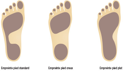
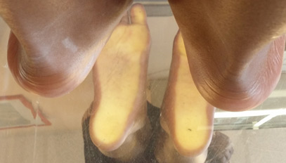
What is Flatfoot?
Flatfoot is a condition where the footprint is widened. Clinically, the inner arch is less pronounced, sometimes even absent. Radiographic assessment shows normal axes, and in advanced cases, there may be a misalignment seen on the profile view, involving the bones of the first ray with the talus, the first cuneiform, and the navicular bone with a downward collapse of the line. In reality, it is mostly the footprint of the foot that determines the definition. To evaluate it, a pedograph or podoscope (see below) must be used. The pedograph allows marking the footprint by passing a layer of ink on a sheet of paper.

What is a Valgus Foot?
The hind foot is “slightly”. With respect to the axis of the leg and is directed towards. The external, lateral side of 5 to 7 °, is the physiological valgus. When this angle is equal or> 10 °, we speak of excess valgus of the hindfoot. This gives the foot an aspect “lying inward, towards the medial side of the foot”. In children, an exaggerated valgus may be corrected with growth; it is the persistence of this angle> 10° which can sign a pathology.
What is a Flat Feet Valgus F.F.V. ?
When the exaggerated valgus is associated with a flat footprint, it is referred to as FFV. Because one can encounter a hollow valgus foot, especially in the “Pes Pronatus”, or more simply when the forefoot is in pronation, ie discreet internal (medial) rotation around the big toe.
Flat feet Q & A
How to treat the flat foot ?
The flat foot is well tolerated. It does not require treatment more often. When this is indicated, it is essentially medical:
- Footwear,
- Insoles,
- Exercises.
With advice for adapting the footwear, with symptomatic care. Wearing plantar orthoses often solves the problem of cramps and pain. Exercises in children and adolescents are very useful. In some cases it is necessary to wear orthopedic shoes, made to measure.
Is it necessary to operate the flat foot?
No, the surgical treatment is very rare, exceptional. It is aimed at forms of neurological etiology and / or severe, disabling forms and in adults when osteoarthritis becomes very painful and the footwear is very difficult or even impossible, or when there is a total eruption of The ark with difficult to walking. Several techniques exist then of which:
- Arthrorysis: temporary blockage of a joint without destruction of the articular surfaces,
- Arthrodesis: also known as double arthrodesis because it blocks both articular surfaces of the calcaneus (see ankle),
- Osteotomy of varus calcaneus: Section of the bone in order to straighten an axis, is done with the surgical saw, or fragilizing the bone by perforations all around the bone, in postage stamp making the correction of bone possible by A simple manual tightening manual. It is a sort of “fracture” for therapeutic purposes.
What are the signs of the flat foot, and those of valgus foot?
In children, adolescents and Young adult, there are no or few symptoms, the foot is very often asymptomatic. At the most, wear and / or deformation characteristic of the shoes can be noted. Sometimes it is the family that finds that the child walks with “feet on or inside”. In adulthood, if the deformity is significant, there may be pain, a deterioration of the mid-foot joints, with the formation of an osteoarthritis of the mid-tarsal articulations , which is associated with an insufficiency of the posterior hamstrings with stretching or a tendon tear or sometimes with a partial or total rupture of their tendons, leading to an arcing of the arch with subluxation of the hind foot.
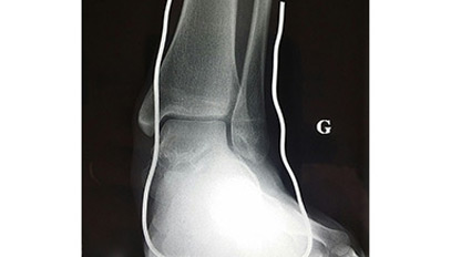
Flat foot, radio of the face of the ankle with rim back foot incidence of Méary
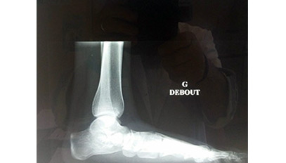
Flat Foot subsidence on profile of the 1st Radius Rupture line of Schade
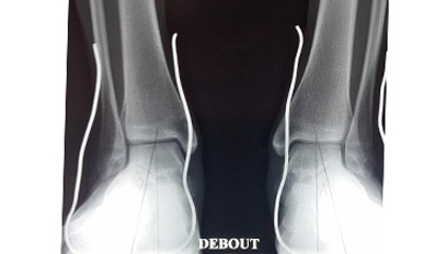
Flat feet Incidence of Méary Valgus 12° and 13° normally 5° to 7°
Pied plat et pied plat valgus :
terminologie
Internal arch: it is the arch that draws foot on its medial or internal edge.
Anterior arch: this is the aspect that the forefoot makes when viewed in a diving form.
Arthrorisis: temporary blockage of a joint such as Grice’s operation.
Arthodesis: definitive blocking of a joint with destruction of the cartilaginous surfaces and bone fusion between the 2 bone parts constituting the joint.
Plantar callus: Thickening of the skin, indicating an area of hyper-pressure.
Podoscopic examination: is an examination that is made standing on a podoscope that shows by fluorescence on the pressure areas and the shape of the imprint. This review is qualitative.
Baro-podometric examination: this is a podoscope equipped with an electronic platform ( having electronic gauges), which make it possible to measure the pressure / cm2 and which by software would visualize this pressure, on a screen, by a chart or a color like a blue and green for light or even normal pressures, yellow and red for areas of high pressure, which reveal points of hyper pressure. If these red zones sits at the pain points, this would explain that there is a mechanical reason for the pain. It’s a quantitative review.
Exostosis: Bony protrusion, at a distance from the joint, its formation is related to the excessive friction of the bone against a rather rigid body, the shoe for example.
Podograph: is a device that allows the taking of the footprint with a sheet and an ink.
Pes pronatus: foot which rotates inwards around an axis passing through the 2nd metartasian and the rear foot.
Podoscope: is a device that allows the taking of the footprint on a mirror, it allows a qualitative examination.
Osteophitis: bone formation around the joint, linked to the development of articular osteoarthritis. It is visible on the x-ray.
Osteotomy: section of the bone to straighten an axis, is done with a surgical saw, or by weakening the bone by perforations all around the bone, in a postage stamp making bone correction possible by a Simple manual tensioning effort. It is a sort of “fracture” for therapeutic purposes.
Plantar vault: this is the shape of the internal arch, the one seen on the medial edge of the foot.
