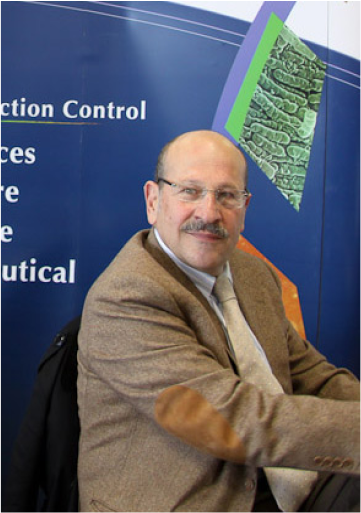
Dr Richard Borgi
Member of the French College of Orthopedic Surgeons
Qualified in Orthopedic Surgery for about 30 years, Dr. R. Borgi has been practicing ankle and foot surgery for more than a decade. He has published several articles and numerous books, one of which was prefaced by Professor R. Merle d’Aubigné and the other by Dr. J. Judet.
- C.E.S: Certificate of Special Studies in Sport Medicine and Biology and kinesiology
- Higher Education: Certificate of Higher Education in Biomechanics of the Musculoskeletal System and Kinesiology.
Expert in Biomechanics
and Surgery
After numerous patents in France and the United States, he participated in research and taught Biomechanics for several years in various universities. He has participated in transfers, to the industry, of innovative technologies in the field of Medical Device. He practices percutaneous and minimally invasive surgery of the foot and ankle the Yonne, at the Polyclinic Sainte Marguerite in Auxerre and in the Loiret at the Clinic of Montargis.
He has published 3 books in the field of Orthopedics, Traumatology and Biomechanics:
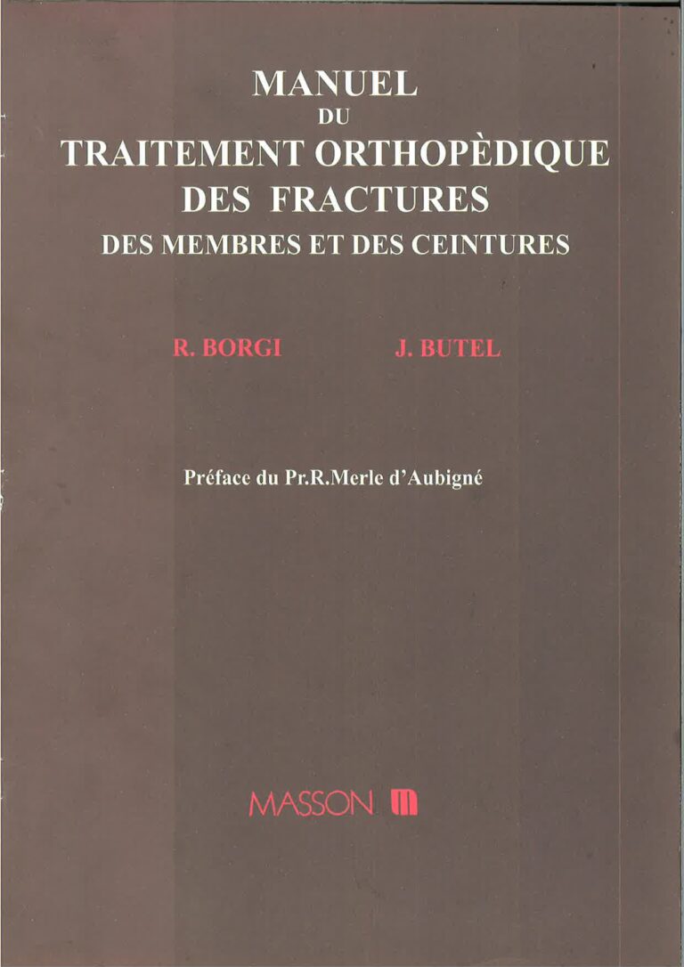
- Manual of orthopedic treatment of fractures of limbs and belts, with J. Butel Préface Pr Merle d’Aubigné, editions Masson (Read the Pr Merle d’Aubigné preface )
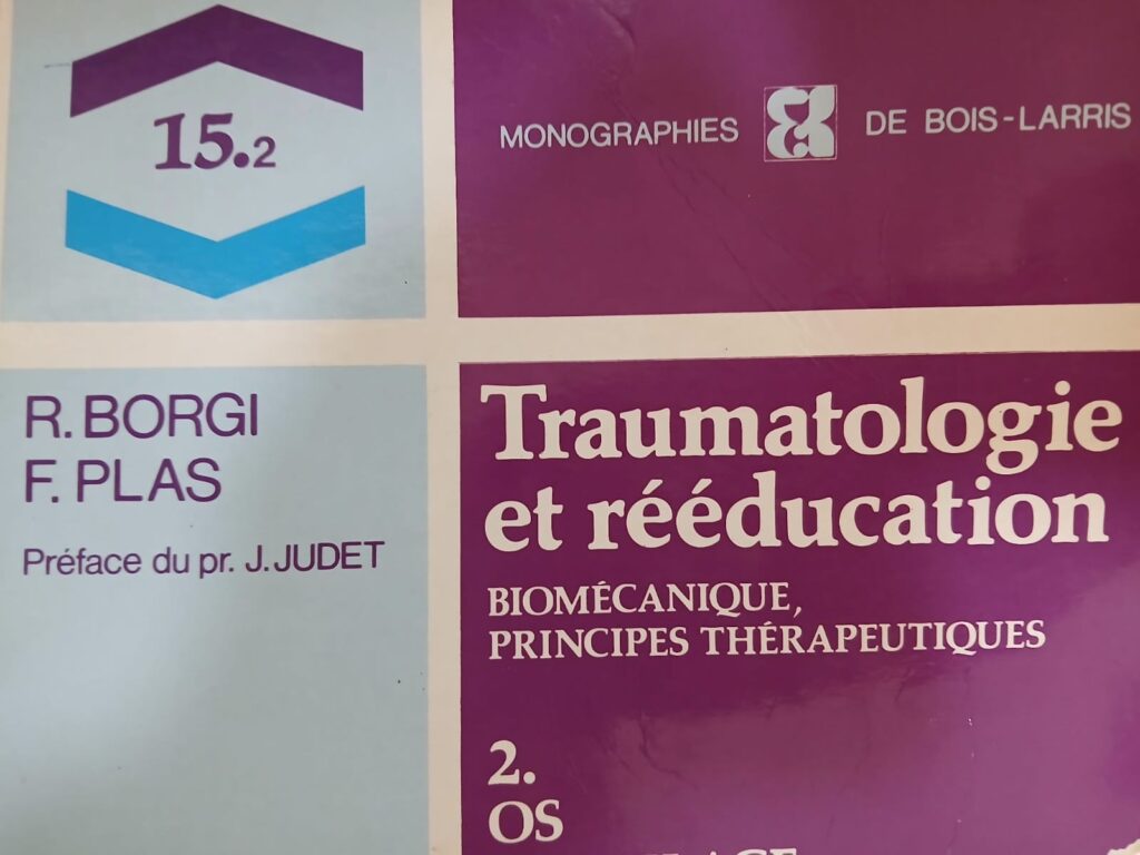
- Traumatology and Reeducation, Volume I, Biomechanical Principles with F. Plas, Preface by Jean Judet to the monographs of Bois Larris, éditions Masson
- Traumatology and Rehabilitation, Volume II, Biomechanical Principles with F. Plas, Preface Dr. Jean Judet in the monographs of Bois Larris, éditions Masson
- Chir. Ortho. : Goal care- editions Mooring
And contributed to the writing of the 2 works:
- Primary osteochondritis of the hip: with Rémy Koehler and Raphael Seringe: SOFCOT Teaching Papers, published by l’Expansion,
- Surgery with A. Kara published by Lamarre
Manual of Orthopedic Treatment of Limb and Girdle Fractures
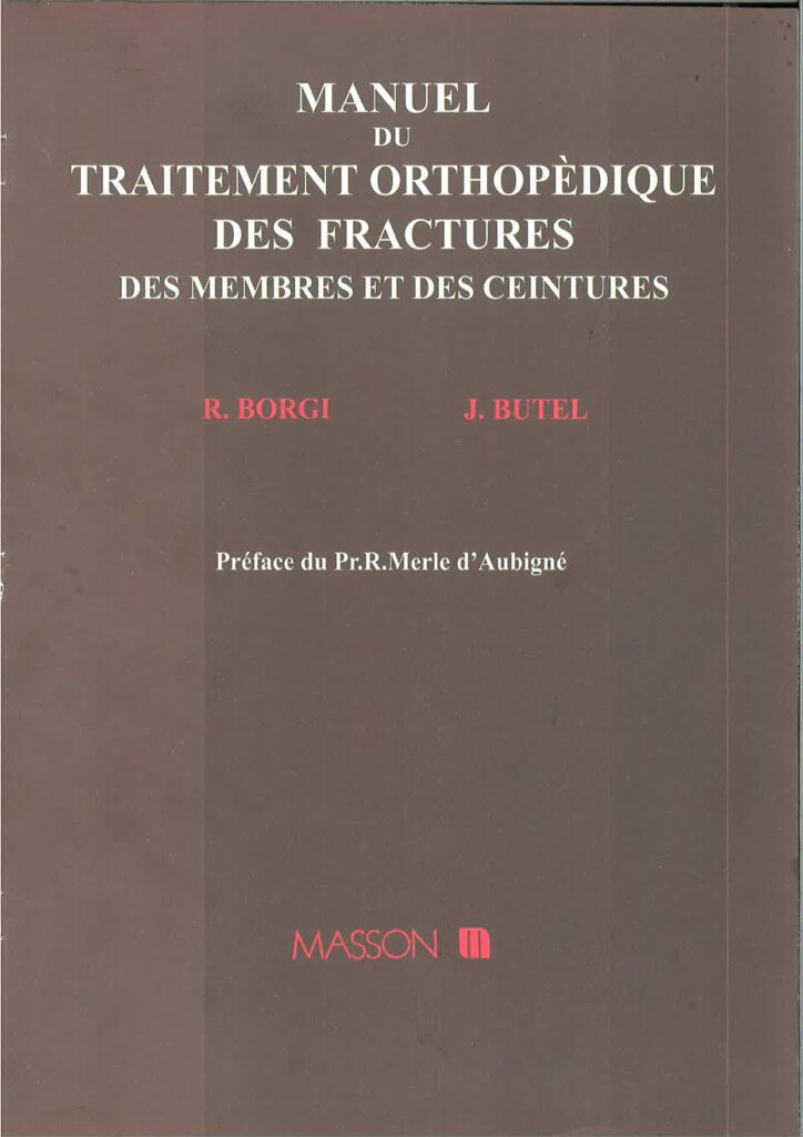
R. Borgi et J. Butel
Ortho, the Orthopedic and Trauma Nurse
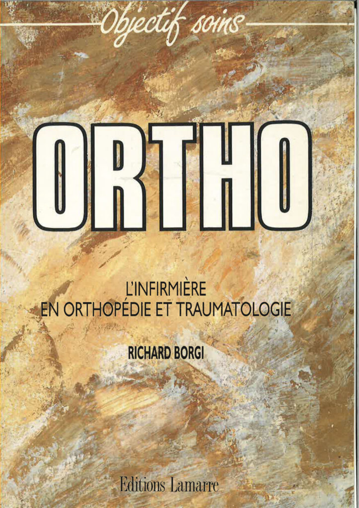
Richard Borgi
Primary Osteochondritis of the Hip
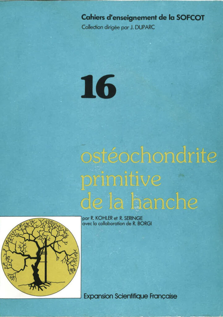
par R. Kohler et R. Seringe avec la collaboration de R. Borgi
Preface by Professor Merle d'Aubigné
In this respect, their book (R. Borgi & J. Butel, authors of the Manual of Orthopedic Treatment of Fractures) is timely. It will be of great service to those who have to treat many fractures, especially where economic constraints are significant — something that may well occur even in “developed” countries.
R. Merle d’Aubigné
Yes, it is time that heals fractures—not thanks to the surgeon or to this or that method, but thanks to that physiological biomechanics which my admiration would call divine, were I not afraid of being accused of teleology: a periosteal sheath which, even when lifted and torn, is sealed enough to keep the spindle-shaped hematoma pressurized around the break; cells contained in this hematoma transform into osteoblasts in the immobile zones at the ends of the spindle, and into cartilage cells in the still-mobile zone:
This cartilaginous callus is solid enough by its shape to hold the fragments in place, yet supple enough not to tear; then comes the invasion of this cartilage by blood vessels, and its ossification forms a provisional callus—still spindle-shaped, therefore solid, but not yet plastic enough to respond to mechanical stress; eventually, these stresses guide the construction of trabeculae and reconstitute a structure adapted to its function, with resorption of the now-useless part of the provisional callus, and often (especially during growth) a correction of deformity.
This admirable process, which allows a dog to heal from a fracture simply by walking on three legs, should make surgeons more humble and prevent them from calling any visible callus on an X-ray “malunited,” like a keloid scar, and from boasting—through a mechanicist distortion quickly refuted by recurrent fractures—about the perfection of the “autogenous weld” they claim to create under their more or less compressive screwed plate.
Very recently, I was asked to testify in a lawsuit brought by a young woman against her surgeon. At age 23, she had sustained a closed fracture of the femoral shaft. After eleven operations and ten years of indescribable suffering and expenses, she was being offered a high-thigh amputation. This took place in a major center in North America.
I felt justified in replying that there was not enough gold in Fort Knox to compensate the young woman for the loss of the ten most beautiful years of her life and ultimately, of one of her lower limbs.
At the opposite end of the long line of witnesses accusing fracture surgery, I might submit this personal case: in a curious accident, a 21-year-old man sustains two identical fractures on both legs—transverse fractures of the mid-shaft of the tibia and fibula. Both are anatomically reduced, realigned, and immobilized in a cast.
But in one case, the reduction was only possible with a small surgical opening, though no osteosynthesis was performed. On the closed side, the tibia consolidated in fifty days; the open fracture site was still mobile at 120 days and had to be treated with a screw fixation graft, successfully.
This case gave me, nearly forty years ago, a conviction that all subsequent observations have confirmed: opening a fracture site—even without the introduction of foreign bodies, even without infection—creates a handicap that must be offset by a solid and lasting fixation, and justified by real benefits in quality of reduction, patient comfort, and, at best, shorter hospitalization.
To the question posed by a participant at the famous Davos osteosynthesis course—”Which fractures can still be treated without surgery?”—one of the instructors gave a flawless answer:
“All those that heal as well without surgery.”
But sadly, most professors were no longer confident in healing through closed methods, and their students were not about to become so…
Because osteosynthesis techniques are so pleasant, their instruments so fascinating, the perfection of radiographic images so satisfying to show the patient, these methods—vigorously promoted by inventors, manufacturers, and vendors—have spread like wildfire, despite their cost, despite the risks, and far beyond reasonable indications. And those who have embarked down this path will find it very difficult to turn back, because they often lack the knowledge, the equipment, and the confidence necessary for the so-called orthopedic treatment of fractures. This treatment is the safest—but it is also the hardest for the surgeon: instead of being resolved in a single operation, it requires many actions with no prestige, and above all, sustained and prolonged attention—therefore, real organization.
While reading their book, I do, however, have two regrets.
The first is that they did not choose, instead of orthopedic treatment, the title closed fracture management: this would have allowed them to include closed intramedullary nailing of the femur and tibia, which—when performed with a closed technique—are the gold standard methods. The exclusion of pinning through the fracture site, which is not highly recommended, would not have been a great loss.
My second regret is that they did not more clearly distinguish between cases where the choice between orthopedic (non-surgical) and surgical treatment is left to the surgeon, based on personal preference or that of the patient, and cases where one method is clearly indicated over the other.
Among the former, I would have included—alongside simple diaphyseal fractures—fractures of the trochanteric region, which they choose to operate on, but which can perfectly well be treated by continuous traction.
By contrast, I believe surgical intervention is clearly indicated in all articular fractures, when it offers the possibility of accurate reconstruction of the joint surfaces—in particular, at the lower ends of the humerus and femur, the upper and lower ends of the tibia, but also the talus and sometimes the calcaneus.
In all cases where accurate reconstruction of joint surfaces is deemed impossible or proves to be so, early mobilization becomes the main imperative, with or without continuous traction (e.g., calcaneus, comminuted fractures of the tibial tuberosities, or the upper end of the humerus), or following excision (e.g., patella, radial head in adults).
Finally, why not clearly state that surgery is as rarely indicated in clavicle fractures as in rib fractures, and never in scapular fractures? But the authors were wise to avoid taking a firm stance on the difficult dilemma of acetabular fractures and complex fractures of the distal femur—which are perilously threatened on the surgical side by inefficacy and worsening, and on the conservative side by poor functional outcomes.
Preface by Professor Merle d'Aubigné
The value of this book is considerable, as their entire therapeutic approach and all their rehabilitation guidelines are based on anatomical, mechanical, and biological data. It is an impressive achievement to have been able, in each chapter, to first detail the fundamental principles and then draw practical conclusions.
A book like this cannot be summarized; it must be read. It is not merely a summary or compilation, but in many chapters presents original expositions. Illustrations support the texts. Rehabilitation doctors, and even general practitioners, will, in my opinion, find in this work a wealth of material for reflection.
J. Judet
Member of the Academy of Surgery, Professor at the College of Medicine of the Hospitals of Paris.
Maison de santé pluridisciplinaire
5 promenade des Champs Plaisants, 89100 SENS
06 34 40 62 29
3C : Centre de Consultations Chirurgicales
7 Av De La Fontaine Ste Marguerite, 89000 AUXERRE
06 34 40 62 29
The conditions described and the treatments mentioned in various sections of this website are for informational purposes only. Only a medical professional can give you advice, and a specialist surgeon can explain your condition during a consultation and, if appropriate, choose with you the most suitable treatment for your body and your specific case.
It is currently acknowledged that smoking increases the risk (both skin and bone complications) in foot surgery by a factor of 2 to 10. Smoking disrupts the natural vascular healing process of wounds (i.e., healing), which can lead to wound dehiscence or even skin necrosis, and is also associated with delayed or failed bone healing.
This is why it is strongly recommended to stop smoking 6 weeks before and 6 weeks after the surgery.
* These risks include: a higher rate of postoperative complications, particularly with impaired healing of surgical wounds (incisions). Healing is the natural process by which surgical incisions close and recover—a process that tobacco hinders or disrupts. If postoperative swelling is also present (e.g., varicose veins, spider veins, venous disorders—especially any condition affecting venous blood return), the risk associated with smoking is further worsened by blood stagnation in the operated areas and the appearance of prolonged edema, which—by putting tension on the incision sutures—slows down the normal healing process.
Articles in scientific journals:
- Review of Orthopedic Surgery,
- R.C.O. Acta Orthopédica Belgica,
- Journal of Surgery,
- Journal of Sports Medicine,
- Annals of Physiotherapy Review of Pediatric Surgery
Articles with one or more co-authors:
- Pr Philippe Merloz (Grenoble)
- Pr Bernard Moyen (Lyon)
- Pr François Bonnel (Montpellier)
- Pr Raphaêl Seringe (Paris)
- Pr Jean Butel (Grenoble)
- Pr Dominique Saragaglia (Grenoble)
- Pr Claude Faure (Grenoble)
- Dr Pascal Oberlin (Grenoble)
- Dr Rémy Boehler (Lyon)
- Dr Pierre Mignot (Valence)
- Dr D. Finidori (Paris)
Of Those:
- The notion of creep in biomechanics,
- Natural History of the Fracture,
- Mechanical and biological aspects of the use of the osteosynthesis plate…
- Femoral neck osteosynthesis: comparative study of mechanical stability,
- Achilles tendon rupture: Orthopedic or surgical treatment?
- Total patellectomy,
- The treatment of retro-spinal disintegration of the LCP, Trickey’s Way,
- The orthopedic treatment of the fracture of the carpal scaphoid,
- Clinical case: loss of diaphyseal bone substance, radial in children,
- The Latarjet operation,
- Blount’s disease,
- Centro-medullary nailing fasciculated: about 275 cases,